 English
 Español
 Português
 русский
 Français
 日本語
日本語 Deutsch
 tiếng Việt
 Italiano
 Nederlands
 ภาษาไทย
 Polski
 한국어
한국어 Svenska
 magyar
 Malay
Malay বাংলা ভাষার
 Dansk
Dansk Suomi
Suomi हिन्दी

Products


Product Description
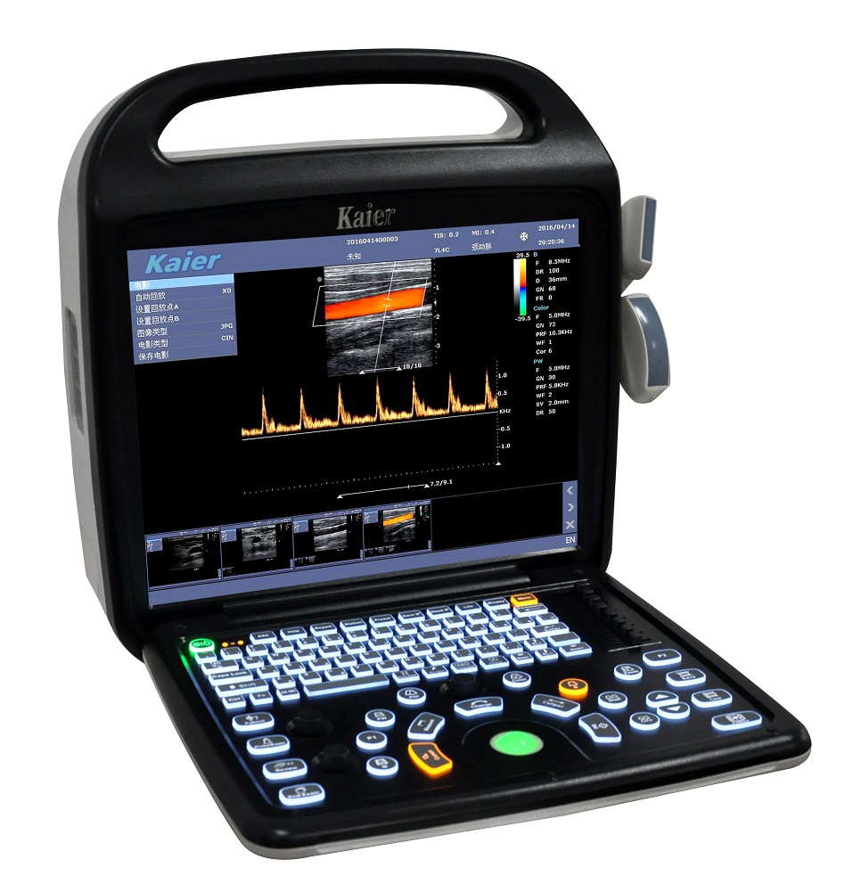
Color Doppler Ultrasound Imaging System
1. General Information
A brand-new ultrasound diagnostic platform with Innovations in areas of digital electronics achieve a new level of ultrasound diagnostic precision and higher diagnostic confidence.
A revolutionary workflow control is provided with the user-centric architecture of the new software platform.
2. Main technical parameters and functions
2.1 Technical platform
linux +ARM+FPGA
2.2 Channels and elements
Number of physical channels: ≥64
Number of probe array element number: ≥128
2.3 Size and weight
Machine size:40cm(front-back)*38cm(left-right)*35cm(height)
Packaging size:52cm(length) * 52cm(width)* 45cm(height)
machine weight: 6.5kg (with no probe)
total weight(include:machine、wooden box、2 probes): 13kg
2.4 Monitor
15-inch, high resolution, progressive scan, Wide Angle of view
Resolution:1024*768 pixels
Image display area is 640*480
2.5 Hard disk
Internal 500GB hard disk for patient database management
Allow storage of patient studies that include images,clips,reports and measurements
2.6 Transducer Ports
Two universal transducer ports
156-pin connection
Unique industrial design provides easy access to all transducer ports
2.7 Probe available
3C6C: Wide convex
7l4C/10L25C: Linear
6C15C/3C20C: Micro convex
6E1C: Endocavity Convex
6I7C: Intrarectal Linear
2P2F: Phased array
2.8 Imaging modes
B-mode: Fundamental and Tissue harmonic imaging
Color Flow Mapping (Color)
Power Doppler Imaging (PDI)
PW Doppler
M-mode
2.9 frequency number
B/M:Fundamental wave,≥3; harmonic wave: ≥2
Color/PDI: ≥2
PW: ≥2
2.10 Cine
B mode: ≥5000 frames
B+Color/B+PDI mode: ≥2500 frames
M、PW: ≥ 190s
2.11 image zoom
available on live, 2B, 4B and reviewed images
up to 10X zoom
2.12 image save
format:
BMP、JPG、FRM(single image);
CIN、AVI(multiple images)
Support DICOM, conform to DICOM3.0 standard
Built in workstation,support patient data search and browse
2.13 language
Support Chinese、English、Spanish、French、German、Czech 、Russian languages.
Can be easily extended to support other languages
2.14 battery
Built in large capacity lithium battery, working condition. Continuous working time ≥1 hours. Screen provides power display information
2.15 Other functions
Comment、BodyMark、Biopsy、Lito, IMT 、report template, support usb mouse ,etc
3. imaging Parameters
3.1 B mode
Up to four frequencies in fundamental imaging
Up to two frequencies in Tissue harmonic imaging (probe dependent)
|
Dynamic range |
0-100% ,5% step |
|
SpeckleReduction |
8 levels(0-7) |
|
ScanDensity |
H,M,L |
|
Gain |
0~100 % ,2% step |
|
TGC |
eight TGC controls |
|
FrameAverage |
8 levels(0-7) |
|
LineAverage |
8 levels(0-7) |
|
Edge Enhance |
8 levels(0-7) |
|
Gray Maps |
15 types(0-14) |
|
Pseudocolor Maps |
7 types(0-6) |
|
Thermal Index |
TIC,TIS,TIB |
|
2B, 4B formats |
/ |
|
Invert (U/D) and transposed (L/R) |
/ |
|
Focus Number |
4 |
|
Focus Depth |
16 levels(depth and probe dependent) |
|
FOV |
5 levels |
|
Image depth up to 35 cm in 0.5~4cm increments (depth dependent) |
|
|
Phase inversion harmonic imaging technique is available for all probes |
|
3.2 Color mode
|
Frequency |
2 levels |
|
Gain |
0~100% ,2% steps |
|
Wall filter |
8 levels(0-7) |
|
Sensitivity |
H,M,L |
|
Flow |
H,M,L |
|
Packet Size1 |
5 levels(0-4) |
|
FrameAverage |
8 levels(0-7) |
|
PostProc |
4 levels(0-3) |
|
Invert |
On/Off |
|
Baseline |
7 levels(0-6) |
|
Color Maps |
4 levels(0-3) |
|
Color/PDI Width |
10%-100%, 10% |
|
Color/PDI Height |
0.5-30cm(probe dependent) |
|
Color/PDI Center Depth |
1-16cm(probe dependent) |
|
Steer |
+/-12°,7°(linear probe) |
3.3 PDI mode
|
Frequency |
2 levels |
|
Gain |
0~100% ,2% steps |
|
Wall filter |
8 levels(0-7) |
|
Sensitivity |
H,M,L |
|
Flow |
H,M,L |
|
Packet Size1 |
5 levels(0-4) |
|
FrameAverage |
8 levels(0-7) |
|
PostProc |
4 levels(0-3) |
|
Invert |
On/Off |
|
Baseline |
7 levels(0-6) |
|
PDI Maps |
2 levels(0-1) |
|
Color/PDI Width |
10%-100%, 10% |
|
Color/PDI Height |
0.5-30cm(probe dependent) |
|
Color/PDI Center Depth |
1-16cm(probe dependent) |
|
Steer |
+/-12°, +/-7°(linear probe) |
3.4 PW mode
|
Frequency |
2 levels |
|
Sweep speed |
5 levels(0-4) |
|
Scale |
16 levels(0-15) (depth and probe dependent) |
|
Scale Unit |
cm/s,KHz |
|
Smooth |
8 levels(0-7) |
|
Pseudocolor Maps |
7 types(0-6) |
|
Dynamic range |
24-100, 2 step |
|
Gain |
0-100%, 2% step |
|
Wall filter |
4 levels(0-3) |
|
Dynamic range |
24-100, 2 step |
|
Gain |
0-100%, 2% step |
|
Wall filter |
4 levels(0-3) |
|
Angle correction |
-89+89,1 step |
|
Gate size |
8 levels(0-7mm) |
|
Wall filter |
5 levels(0-4) |
|
Invert |
On/Off |
|
Baseline |
7 levels |
|
Real-time auto Doppler trace: maximum velocity, mean velocity |
|
3.5 M Mode
|
Frequency |
Up to 3 fundamental and 2 harmonic imaging frequencies |
|
Edge enhance |
8 levels(0-7) |
|
Dynamic range |
0-100%,step 5% |
|
Gain |
0-100,step 2 |
|
Gray Maps |
15 levels(0-14) |
|
Pseudocolor Maps |
7 (0-6) |
|
Sweep speed |
5 levels(0-4) |
3.6 image parameter save and restore
user can press one key to save image parameters in screen
user can press one key to restore image parameters to default status.
4. Ergonomic Design
Frequently used controls centre around the trackball
Control panel is backlighted, waterproof and antisepticised
Two USB port are at the back of the system, which is more convenient for use
5. Exam Modes
Abdomen
Obstetrics
Gynecology
Fetal Heart
Small parts
Urology
Carotid
Thyroid
Breast
Vascular
Kidney
Pediatrics
6. Product configuration
6.1 Standard configuration
Host(Built-in 500G hard disk)
3C6C convex array probe
7L4C linear array probe
User's Manual
Power cable
6.2 Optional Accessories
6E1C Endocavity Convex probe
10L25C linear array probe
6I7C Intrarectal Linear probe
6C15C Micro convex probe
3C20C Micro convex probe
2P2F Phased array probe
USB report printer
B/W or color Video printer
Puncture rack
Foot switch
U disk and USB extension line
Hot Tags:
Product Tag
Send Inquiry
Please feel free to fill your inquiry in the form below. We will reply you in 24 hours.


We use cookies to offer you a better browsing experience, analyze site traffic and personalize content. By using this site, you agree to our use of cookies.
Privacy Policy